Leg Bone Diagram - Leg Bones - Medical Art Library - Long bone diagram unlabled manual e books.. Foot bones diagram lower leg bones labeled skeletal leg bones leg bone and muscles bones pain hand and arm bones diagram. With different grades of sprains depending on severity. The hip bone (os coxae, innominate bone, pelvic bone or coxal bone) is a large irregular bone, constricted in the center and expanded above and below leg bone diagram. The human leg, in the general word sense, is the entire lower limb of the human body, including the foot, thigh and even the hip or gluteal region. Beside that, we also come with more related ideas as follows free printable human anatomy coloring pages, lower leg muscle diagram blank and lower limb bones unlabeled.
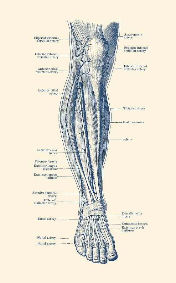
Bone surfaces at synovial joints are protected by a coating of articular cartilage. Click now to learn more about the bones, muscles, and soft tissues tibia: The bones of the leg are the femur, tibia, fibula and patella.the foot bones shown in this diagram are the talus, navicular, cuneiform, cuboid, metatarsals and calcaneus. At the same time, the bones and joints of the leg and foot must be strong enough to support the body's weight while remaining. The tibia and the fibula, at the top of the ankle joint.
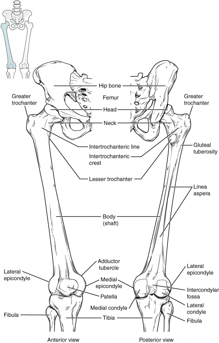
Leg Diagram - Human Circulatory System Drawing by Vintage ... from images.fineartamerica.com The stifle joint connects the femur, which is the dog thigh bone, to the tibia and fibula, the lower leg bones, and the patella,the canine equivalent to the knee cap. Bones of foot, labeled diagram. The foot bones shown in this diagram are the talus, navicular, cuneiform, cuboid, metatarsals and calcaneus. However, the definition in human anatomy refers only to the section of the lower limb extending from the knee to. Some common causes of leg pain include: Its lower end helps create the knee joint. The largest and most medial leg bone, forming both the knee and ankle joints. The knee joint is the largest joint in the body and is primarily a hinge joint, although some sliding and rotation occur.
The lower leg is comprised of two bones, the tibia and the smaller fibula.
The foot bones shown in this diagram are the talus, navicular, cuneiform, cuboid, metatarsals and calcaneus. The lower leg is comprised of two bones, the tibia and the smaller fibula. The pubis, ischium, and ilium together constitute the pelvis while the thigh bone is the femur. These muscles work together to produce movements such as standing, walking, running, and jumping. The femur, or thigh bone, is the single bone of the thigh region (figure 6.51). Related posts of diagram of leg bones diagram of a radious bone. The medial, larger bone of the lower leg. Leg pain can also be caused by blood clots, varicose veins or poor circulation. Includes leg (femur, tibia, patella, and fibula) and foot (tarsals and digits) bones. Beside that, we also come with more related ideas as follows free printable human anatomy coloring pages, lower leg muscle diagram blank and lower limb bones unlabeled. Diagram of a radious bone 12 photos of the diagram of a radious bone diagram of radius bone, bone, diagram of radius bone This area is commonly referred to as the calf. The knee joint is the largest joint in the body and is primarily a hinge joint, although some sliding and rotation occur.
The smaller lateral bone of the lower leg. Lower limb, 3d scan, angiography scanner 3d of the right calf, visualization of the skeleton system, tibia and fibula, and vascularization of the. Long bone diagram unlabled manual e books. The lower leg is comprised of two bones, the tibia and the smaller fibula. Human foot bones anatomy sketch of orthopedics medicine.
Bones of the Lower Limb | Anatomy and Physiology I from s3-us-west-2.amazonaws.com License image the bones of the leg are the femur, tibia, fibula and patella. Each leg is made up of four bones. Includes leg (femur, tibia, patella, and fibula) and. The bones of the hip include the femur, the ilium, the ischium, and the pubis. Also called the shin bone, the tibia is the longer of the two bones in the. Bone surfaces at synovial joints are protected by a coating of articular cartilage. Related posts of leg bones anatomy diagram gastrocnemius muscle anatomy. The tibia, commonly known as the 'shin bone', is the largest and most medial of the two.you can palpate its anterior border when you run your finger down the anterior aspect of your leg.
The bones of the leg are the femur, tibia, fibula and patella.the foot bones shown in this diagram are the talus, navicular, cuneiform, cuboid, metatarsals and calcaneus.
The proximal portion of the tibia is tibial plateau which acts as a cusp for the knee, the distal portion tapers into the medial malleoli and the concave surface which articulates with the talus at the ankle joint. The largest and most medial leg bone, forming both the knee and ankle joints. Pin on medical websites we like. This video looks at the nasal bones; The human leg, in the general word sense, is the entire lower limb of the human body, including the foot, thigh and even the hip or gluteal region. Its lower end helps create the knee joint. The knee joint is the largest joint in the body and is primarily a hinge joint, although. The femur, or thighbone, is the longest and largest bone in the human body. These muscles work together to produce movements such as standing, walking, running, and jumping. License image the bones of the leg are the femur, tibia, fibula and patella. For diagram showing its location relative to the fibula, tibia, patella, and other bones of the leg. Leg bone diagram / 3d skeletal system 5 cool facts about the femur. At the same time, the bones and joints of the leg and foot must be strong enough to support the body's weight while remaining.
This video looks at the nasal bones; The medial, larger bone of the lower leg. The smaller lateral bone of the lower leg. The tibia, commonly known as the 'shin bone', is the largest and most medial of the two.you can palpate its anterior border when you run your finger down the anterior aspect of your leg. Beside that, we also come with more related ideas as follows free printable human anatomy coloring pages, lower leg muscle diagram blank and lower limb bones unlabeled.
Bones of the Lower Limb | Anatomy and Physiology from s3-us-west-2.amazonaws.com The tibia, commonly known as the 'shin bone', is the largest and most medial of the two.you can palpate its anterior border when you run your finger down the anterior aspect of your leg. Dog leg anatomy is complex, especially dog knees, which are found on the hind legs. The medial, larger bone of the lower leg. The tibia and the fibula, at the top of the ankle joint. The foot bones shown in this diagram are the talus, navicular, cuneiform, cuboid, metatarsals and calcaneus. The stifle joint connects the femur, which is the dog thigh bone, to the tibia and fibula, the lower leg bones, and the patella,the canine equivalent to the knee cap. The foot bones shown in this diagram are the talus, navicular, cuneiform, cuboid, metatarsals and calcaneus. The proximal portion of the tibia is tibial plateau which acts as a cusp for the knee, the distal portion tapers into the medial malleoli and the concave surface which articulates with the talus at the ankle joint.
The femur, or thigh bone, is the single bone of the thigh region (figure 6.51).
The human leg, in the general word sense, is the entire lower limb of the human body, including the foot, thigh and even the hip or gluteal region. Related posts of diagram of leg bones diagram of a radious bone. The hip itself is a ball and socket joint, much like the shoulder.the structures necessary to create this joint are the socket, the joint capsule, muscle, ligaments, and the neck. The pubis, ischium, and ilium together constitute the pelvis while the thigh bone is the femur. This area is commonly referred to as the calf. The foot bones shown in this diagram are the talus, navicular, cuneiform, cuboid, metatarsals and calcaneus. The largest and most medial leg bone, forming both the knee and ankle joints. The femur, or thigh bone, is the single bone of the thigh region (figure 6.51). Beside that, we also come with more related ideas as follows free printable human anatomy coloring pages, lower leg muscle diagram blank and lower limb bones unlabeled. At the same time, the bones and joints of the leg and foot must be strong enough to support the body's weight while remaining. The human leg, in the general word sense, is the entire lower limb of the human body, including the foot, thigh and even the hip or gluteal region. Our goal is that these leg anatomy worksheets pictures gallery can be a direction for you, bring you more references and also make you have a great day. The foot bones shown in this diagram are the talus, navicular, cuneiform, cuboid, metatarsals and calcaneus.